
Hydrocephalus — sometimes called "water on the brain" — is an abnormal buildup of cerebrospinal fluid inside the brain's ventricles, which raises the pressure inside the skull. In children it can be present from birth or appear later, and in most cases it can be treated successfully if it's caught early.
What is hydrocephalus?
Hydrocephalus happens when the cerebrospinal fluid that surrounds the brain and spinal cord builds up faster than it drains, so one or more of the brain's ventricles expand and pressure rises inside the skull. In infants whose skull bones haven't fused yet, this often shows up as a increase head circumference. In older children and adults, whose skull is already fully formed, the symptoms look quite different.
What are the symptoms in children?
Symptoms depend on the child's age and how quickly the condition develops. In children under two, the main sign is an increase head circumference, while In children whose skull bones have already fused, symptoms can include:
- Unsteady walking or frequent falls
- Recurring headache, especially in the morning
- A sudden squint or a change in eye movement
- Nausea or vomiting
- Urinary incontinence in a child who was previously toilet-trained
- Excessive sleepiness or a decline in school performance
- Change in the conscoius level (Need urgent surgical intervention)
Not all of these appear together, and sometimes the only noticeable sign is a subtle change in gait or vision — which is why parents often delay a visit despite how much it matters.
What causes it?
One of the most common causes of hydrocephalus in children is aqueductal stenosis — a narrowing of the channel connecting the third and fourth ventricles that blocks the normal drainage of cerebrospinal fluid. Other causes include congenital brain malformations, small tumours pressing on the fluid pathway, prior meningitis, or bleeding inside the brain in newborns, particularly premature infants. Pinpointing the exact cause on imaging is what guides the treatment decision, since treatment differs depending on where and why the blockage occurred.
When should you see a doctor immediately?
Seek urgent evaluation if your child develops any of the following: a severe or worsening headache, repeated vomiting especially alongside headache, a sudden squint or double vision, unusual sleepiness or difficulty waking, or a sudden decline in balance or walking, change in the conscoiusness (Late sign and need an urgent surgical intervention). These signs can point to rising pressure inside the skull and call for prompt evaluation, not a wait for the next routine appointment.
How is it diagnosed?
Diagnosis starts with a clinical exam and, in infants, measuring head circumference, followed by an MRI of the brain that shows the exact location and cause of the blockage. In infants whose soft spot (fontanelle) hasn't closed yet, an ultrasound through it can serve as a quick first step before MRI. The MRI itself is what determines whether the case is suitable for endoscopic surgery.
What are the treatment options?
There are two main treatment options for hydrocephalus: a permanent shunt, which drains the excess fluid to another body cavity such as the abdomen, or endoscopic third ventriculostomy (ETV), which creates a new pathway for cerebrospinal fluid without implanting any permanent tube. A shunt plays an important role in many cases, but it requires long-term follow-up and may need to be revised more than once over a child's life. ETV, by contrast, suits only specific cases, and the choice between the two depends on the exact cause and location of the blockage in each child.
When is endoscopic surgery (ETV) the better option?
Aqueductal stenosis is one of the conditions that responds best to endoscopic third ventriculostomy, with a higher success rate than many other causes of hydrocephalus. In these cases, ETV can spare a child the need for a permanent shunt and the long-term follow-up that comes with it. That said, not every case is a candidate for this option — careful evaluation on imaging is what determines this for each child individually.
How is the endoscopic procedure done?
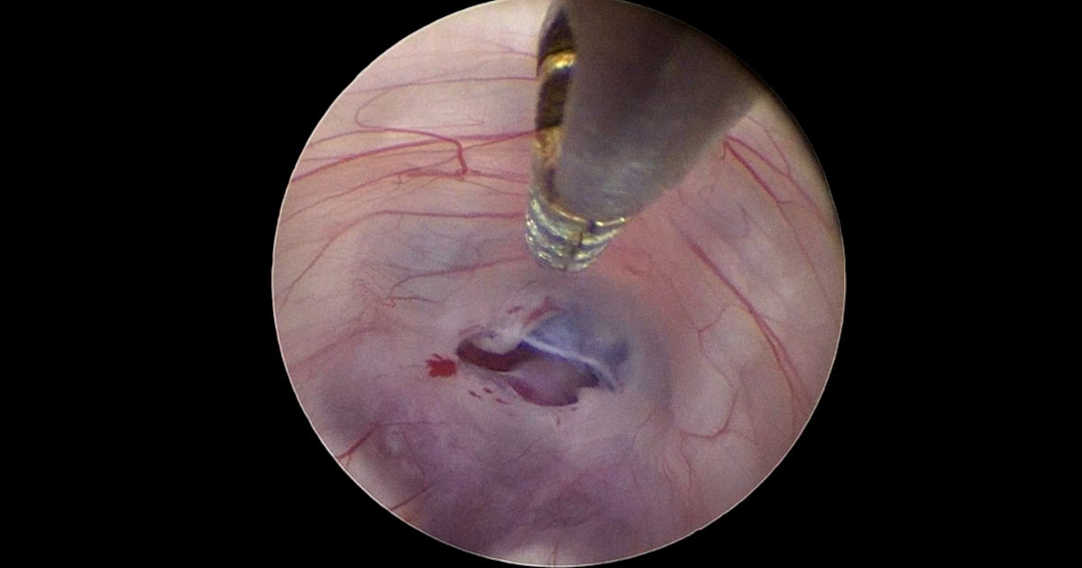
The procedure is done under general anaesthesia. A fine endoscope is passed through a small opening in the skull to reach the third ventricle, where a small opening is made in its floor to create a new pathway that allows cerebrospinal fluid to drain normally, without implanting any foreign material.
How long does recovery take?
The standard hospital stay is one or two days, Recovery time varies from one child to another, but symptoms typically improve gradually over the weeks following surgery, with periodic follow-up imaging to confirm the fluid continues to drain normally over the long term. With a permanent shunt, follow-up tends to run longer, since the shunt may need adjustment or replacement as the child grows.
Can hydrocephalus come back after treatment?
Yes, regardless of which treatment was used. After endoscopic third ventriculostomy, the opening created in the ventricle floor can narrow again months or years later, bringing symptoms back and requiring a new evaluation and possibly another procedure. After a permanent shunt, a blockage or mechanical failure can occur and require replacement. This is why periodic follow-up imaging remains part of the treatment itself, even once a child has fully improved — not a formality to skip.
Book an appointment
If you notice any of these signs in your child, early evaluation makes a real difference. Visit our contact page to book an appointment, or read more about neuroendoscopy and the story of a child treated this way.